

Loading…
Loading…
Loading…
OCR GCSE Combined Science B · J260
OCR J260 · Transport Systems and Exchange Surfaces Check the specification (PDF) (opens in a new tab)
The circulatory system consists of the heart, blood and a network of blood vessels. The heart supplies the pumping force, the vessels provide routes, and the blood carries substances between organs and cells. This connects the places where substances enter or leave the body with the tissues that need them.
In the gaseous exchange system, oxygen diffuses from the alveoli in the lungs into the blood. Blood delivers it to cells for aerobic respiration. Carbon dioxide produced by respiration travels in the opposite direction: from cells into the blood, then to the lungs, where it diffuses into the alveoli and is breathed out.
In the digestive system, digestion breaks food down into small, soluble molecules. Molecules such as glucose and amino acids are absorbed into capillaries in the villi of the small intestine. Blood then distributes them to cells for reactions such as respiration and building proteins.
The circulatory system also connects to the excretory system, which removes metabolic waste. Urea is made in the liver when excess amino acids are broken down. Blood carries it to the kidneys, where it is removed from the blood and leaves the body in urine. Water is transported in the blood too.
Humans have a double circulation. One circuit carries blood between the heart and lungs; the other carries blood between the heart and the rest of the body. Blood therefore passes through the heart twice on a complete journey around both circuits.
The right side of the heart receives deoxygenated blood, which has a lower oxygen content, from the body and pumps it to the lungs. The left side receives oxygenated blood, which has a higher oxygen content, from the lungs and pumps it around the body. Deoxygenated blood is not completely without oxygen.
Blood loses pressure as it flows through the lung capillaries. Returning it to the heart allows it to be pumped again at higher pressure before travelling around the body, helping to supply tissues efficiently.
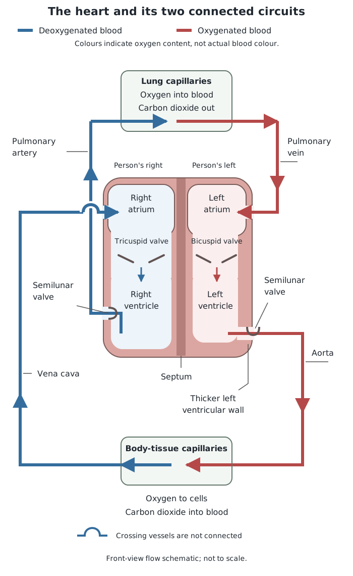
The right side pumps blood to the lungs; the left side pumps blood around the body. Arrows show the direction of flow.
The heart has four chambers. The two upper chambers, the atria, receive blood. The two lower chambers, the ventricles, pump blood out.
Follow a portion of blood through this route:
This describes a continuous route, not six separate beats. Both atria contract together, and then both ventricles contract together. A dividing wall called the septum keeps blood in the right and left sides separate, preventing oxygenated and deoxygenated blood from mixing.
The heart wall contains cardiac muscle. Its repeated contractions generate pressure that moves blood. Cardiac muscle cells contain many mitochondria to support aerobic respiration, supplying the energy needed for continuous contractions. Coronary arteries supply the heart muscle itself with oxygen and glucose.
The atria only push blood into the nearby ventricles, so their walls are relatively thin. The ventricles have thicker muscular walls because they must pump blood out of the heart. The left ventricular wall is thicker than the right ventricular wall: it generates the higher pressure needed to move blood around the whole body, whereas the right ventricle pumps blood to the nearby lungs at lower pressure.
Valves make this pumping effective by ensuring one-way flow. The tricuspid and bicuspid valves lie between atria and ventricles and prevent blood returning to the atria when the ventricles contract. The semilunar valves prevent blood returning from the pulmonary artery and aorta when the ventricles relax.
Arteries carry blood away from the heart. Their thick walls withstand the high pressure produced by ventricular contractions. Thick muscle and elastic layers help control blood flow and maintain pressure: elastic tissue stretches as blood is pumped into an artery and recoils afterwards. Arteries do not have valves along their length.
Veins carry blood towards the heart. By the time blood reaches them, its pressure is much lower. Their walls are thinner, with less muscle and elastic tissue, because they do not have to withstand arterial pressure. Valves prevent the low-pressure blood from flowing backwards.
The pulmonary vessels show why direction, rather than oxygen content, defines a vessel: the pulmonary artery carries deoxygenated blood to the lungs, while the pulmonary vein carries oxygenated blood back to the heart.
Capillaries connect arteries to veins within tissues. Their main role is exchange. Their very thin walls provide a short diffusion distance between blood and cells. Their narrow lumen keeps the blood close to the wall, while extensive capillary networks provide a large surface area. Blood flows more slowly through these networks, allowing time for exchange. Oxygen and nutrients move towards cells, while wastes move into the blood.
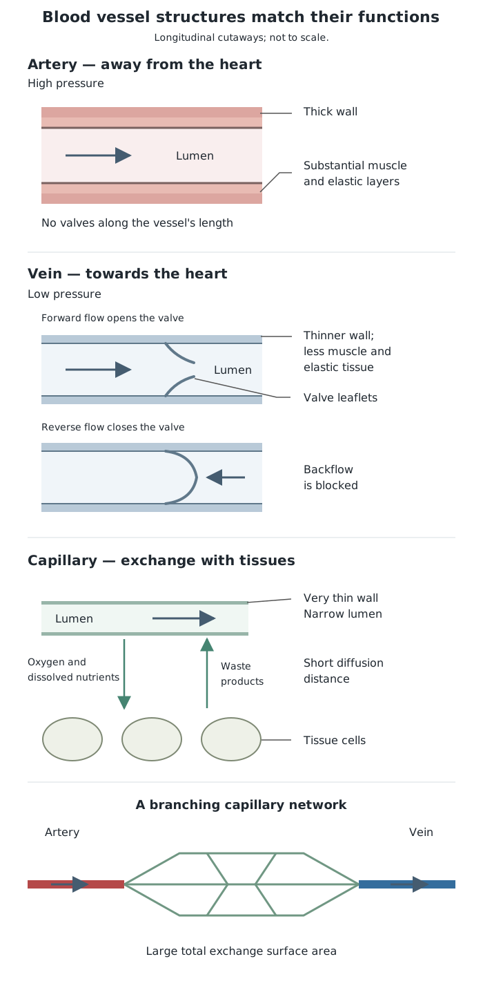
Arteries withstand high pressure, veins prevent backflow at low pressure, and capillaries provide short distances for exchange.
Red blood cells contain haemoglobin, a protein that binds oxygen in the lungs to form oxyhaemoglobin. This binding is reversible, so oxygen can be released in tissues where it is needed.
Their shape is a biconcave disc: both broad surfaces dip inwards. This gives a large surface area relative to their volume for oxygen diffusion and a thin centre that keeps diffusion distances short. Mature red blood cells have no nucleus, leaving more space for haemoglobin. They are also small and flexible, allowing them to pass through narrow capillaries.
Plasma is the liquid part of blood, with blood cells suspended in it. Its water provides a solvent, so many substances can dissolve and be carried as the blood flows.
Plasma transports digested food molecules from the intestine, urea from the liver to the kidneys, and carbon dioxide from tissues towards the lungs. It also carries hormones to their target organs and distributes heat around the body. Red blood cells and plasma therefore have complementary roles: haemoglobin carries oxygen, while plasma carries many other substances in solution.
Double circulation: heart → lungs → heart → body → heart.
Vena cava → right atrium → tricuspid valve → right ventricle → semilunar valve → pulmonary → lungs → pulmonary → left atrium → bicuspid valve → left ventricle → semilunar valve → aorta → body.
Define arteries and veins by the direction of blood flow, not by oxygen content: arteries carry blood away from the heart.
On a front-view heart diagram, the person's right side is on your left. Follow the vessel connections and labels.
Link each adaptation to its function: for example, a thicker left ventricular wall produces higher pressure for circulation around the body.
Distinguish valves from muscle: muscle contractions generate pressure; valves prevent backflow.
Red blood cells carry oxygen using haemoglobin; plasma carries dissolved substances such as urea and digested food molecules.
It can see this note and what your specification asks for.
Answers are free with an account.
1 free note left this week
Get more free notesYour answer sets when this note comes back for review, or marks it complete.
Was this note helpful?
Log in to tell us whether it helped, or to report a problem.
Last updated
Circulatory system
The heart, blood and blood vessels that transport substances around the body.
Double circulation
A circulation in which blood passes through the heart twice during one complete journey through the lungs and the rest of the body.
Cardiac muscle
The specialised muscle forming the heart wall, which contracts repeatedly to pump blood.
Atrium
An upper chamber of the heart that receives blood returning in veins.
Ventricle
A lower chamber of the heart that pumps blood out into an artery.
Valve
A structure that prevents blood flowing backwards.
Artery
A blood vessel that carries blood away from the heart.
Vein
A blood vessel that carries blood towards the heart.
Capillary
A very small, thin-walled blood vessel where substances are exchanged between blood and surrounding tissues.
Lumen
The space inside a blood vessel through which blood flows.
Haemoglobin
A protein in red blood cells that binds oxygen reversibly, allowing oxygen to be picked up in the lungs and released to tissues.
Plasma
The liquid component of blood in which blood cells are suspended and many transported substances are dissolved.
Put your knowledge into practice — try past paper questions for Combined Science B
Circulatory system
The heart, blood and blood vessels that transport substances around the body.
Double circulation
A circulation in which blood passes through the heart twice during one complete journey through the lungs and the rest of the body.
Cardiac muscle
The specialised muscle forming the heart wall, which contracts repeatedly to pump blood.
Atrium
An upper chamber of the heart that receives blood returning in veins.
Ventricle
A lower chamber of the heart that pumps blood out into an artery.
Valve
A structure that prevents blood flowing backwards.
Artery
A blood vessel that carries blood away from the heart.
Vein
A blood vessel that carries blood towards the heart.
Capillary
A very small, thin-walled blood vessel where substances are exchanged between blood and surrounding tissues.
Lumen
The space inside a blood vessel through which blood flows.
Haemoglobin
A protein in red blood cells that binds oxygen reversibly, allowing oxygen to be picked up in the lungs and released to tissues.
Plasma
The liquid component of blood in which blood cells are suspended and many transported substances are dissolved.
| Vessel | Function and adaptation |
|---|---|
| Artery | Away from heart; thick muscular, elastic walls withstand high pressure. |
| Vein | Towards heart; thinner walls suit low pressure; valves prevent backflow. |
| Capillary | Exchange; thin walls, narrow lumen, extensive networks and slower flow. |
Pulmonary artery: deoxygenated blood. Pulmonary vein: oxygenated blood.