

Loading…
Loading…
Loading…
OCR GCSE Combined Science B · J260
OCR J260 · Cell Structures, DNA, and Protein Synthesis Check the specification (PDF) (opens in a new tab)
Cells are the basic units of living organisms. Their genetic material is DNA, which carries instructions controlling how they develop and function. These instructions allow cells to make proteins, including enzymes that control chemical reactions. This connects genetic material to everyday cell activities such as growth and using nutrients.
Plant and animal cells are eukaryotic cells. Their main genetic material is enclosed in a nucleus, where DNA is packaged into several long, linear chromosomes. The nucleus controls cell activities because it contains these instructions—not because it physically carries out every process itself.
Other structures carry out particular jobs. Many chemical reactions happen in the cytoplasm, the cell membrane controls movement of substances into and out of the cell, and ribosomes make proteins. The genetic instructions and these working structures therefore act together.
Bacteria are prokaryotic organisms. Their cells are generally much smaller than plant and animal cells and have no nucleus. Instead, their main DNA forms a long circular chromosome in the cytoplasm. This DNA still controls cell activities and provides instructions needed when the cell reproduces.
Bacteria may also contain plasmids: small circles of extra DNA separate from the main chromosome. Plasmids carry additional genetic instructions. For example, a plasmid may carry instructions that give a bacterium resistance to an antibiotic, allowing it to survive exposure to that antibiotic. Plasmids can be transferred between bacteria, so useful instructions can spread.
The important distinction is where the DNA is kept and how it is organised. Both eukaryotic and prokaryotic cells have genetic material; only eukaryotic cells enclose their main genetic material in a nucleus.
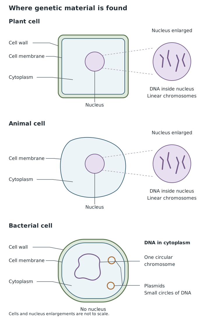
Plant and animal cells enclose their main DNA in a nucleus; bacteria have a circular chromosome in the cytoplasm and may also have plasmids. Cells are not shown to scale.
A light microscope passes visible light through a specimen and uses lenses to produce an enlarged image. The objective lens sits above the specimen; the eyepiece lens is the lens you look through. Both magnify the image.
The slide rests on the stage, with the specimen above a hole through which light passes. Stage clips hold it in place. The coarse focus makes large adjustments to bring cells roughly into focus, while the fine focus makes small adjustments to sharpen their outlines. The light source and diaphragm let you adjust illumination and contrast.
A specimen must be thin enough for light to pass through it. Overlapping layers make structures difficult to distinguish. Many cells are almost transparent, so adding a stain makes structures such as the nucleus stand out against their surroundings.
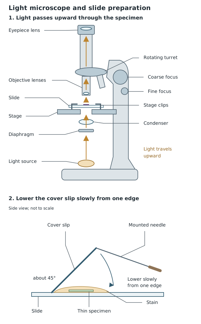
Light passes through a thin specimen before reaching the lenses. Rest one edge of the cover slip on the slide beside the drop, then lower it slowly to help prevent trapped air bubbles.
For this investigation, use a light microscope, clean slides and cover slips, forceps, a mounted needle, dropping pipettes, iodine solution, methylene blue, filter paper and a sterile cotton bud. Onion epidermis provides a plant specimen, while cheek cells provide an animal specimen.
Onion epidermis: use forceps to peel a small piece of the thin skin from the inner surface of a fleshy onion layer. Lay it flat in the centre of a slide, removing folds so that cells do not overlap. Add a drop of iodine solution to improve contrast.
Cheek cells: gently rub the inside of your own cheek with a sterile cotton bud. Smear the collected cells thinly across a separate slide, then put the used bud straight into disinfectant. Add a drop of methylene blue to make the nuclei easier to see.
For either specimen, rest one edge of a cover slip beside the drop, holding it at roughly with a mounted needle. Lower it slowly so that the liquid spreads beneath it without trapping air. The cover slip flattens and protects the specimen and keeps liquid away from the objective lens. Use filter paper at its edge to absorb surplus liquid.
Wear eye protection and avoid getting stains on skin or clothing. Do not ingest stains or share cotton buds. Handle glass and the mounted needle carefully. If cutting onion tissue, cut on a tile away from your fingers. Carry the microscope with one hand supporting its base and the other holding its arm.
The view is ready to record when individual cells have sharp outlines and their stained nuclei can be distinguished. If the image remains blurred, return to low power and check the focus, specimen thickness and slide preparation.
Total magnification is found by multiplying the two lens magnifications:
For example, a ×10 eyepiece and a ×40 objective produce ×400 magnification. The image is enlarged 400 times; this does not mean every tiny structure becomes visible.
Onion epidermis cells usually look like closely packed, brick-shaped compartments. Their clear boundaries are cell walls, and iodine helps reveal the nuclei within them. This onion bulb tissue does not contain chloroplasts, so a plant cell need not show every structure in a general textbook diagram.
Cheek cells are flatter, irregularly rounded cells that may lie separately or overlap. Methylene blue reveals a darker nucleus within the cytoplasm. They have a cell membrane but no cell wall.
Seeing nuclei in both specimens supports their classification as eukaryotic cells. However, you cannot make out individual DNA molecules or plasmids with a school light microscope. A diagram of genetic material explains its organisation; it is not a picture of everything this practical reveals.
Record a representative group of cells using clear, single pencil lines without shading. Add a descriptive title, label visible structures with straight ruled lines and record the lens magnifications. The drawing should show your observations rather than an idealised cell.
When comparing genetic material, state both its location and its form: chromosomes inside a nucleus in plant and animal cells; a circular chromosome in the cytoplasm of a bacterial cell, sometimes with plasmids.
A bacterium has no nucleus, but it still has DNA that controls its activities.
Explain practical choices: a thin specimen lets light through, a stain improves contrast, and starting at low power makes cells easier to find.
Use only fine focus at high power to avoid driving the objective lens into the cover slip.
Draw and label only structures you actually observe. Onion bulb epidermis cells do not contain chloroplasts.
Total microscope magnification is eyepiece magnification multiplied by objective magnification. Magnification has no unit.
It can see this note and what your specification asks for.
Answers are free with an account.
1 free note left this week
Get more free notesYour answer sets when this note comes back for review, or marks it complete.
Was this note helpful?
Log in to tell us whether it helped, or to report a problem.
Last updated
Genetic material
The DNA that carries instructions controlling how a cell or organism develops and functions.
Eukaryotic cell
A cell with a nucleus containing its main genetic material. Plant and animal cells are eukaryotic.
Prokaryotic cell
A cell without a nucleus, with its main genetic material in the cytoplasm. Bacteria are prokaryotic.
Nucleus
A structure in a eukaryotic cell that contains chromosomes made of DNA, whose instructions control the cell's activities.
Chromosome
A long DNA molecule carrying genetic instructions.
Plasmid
A small circular DNA molecule that may occur in a bacterial cell, separate from its main chromosome.
Cytoplasm
The gel-like material inside a cell where many chemical reactions take place.
Light microscope
An instrument that uses visible light and lenses to produce a magnified image of a specimen.
Specimen
The biological material being examined, such as a thin layer of cells mounted on a microscope slide.
Stain
A coloured chemical used to increase contrast so that cell structures are easier to distinguish.
Magnification
The number of times larger an image is than the actual object.
Put your knowledge into practice — try past paper questions for Combined Science B
Genetic material
The DNA that carries instructions controlling how a cell or organism develops and functions.
Eukaryotic cell
A cell with a nucleus containing its main genetic material. Plant and animal cells are eukaryotic.
Prokaryotic cell
A cell without a nucleus, with its main genetic material in the cytoplasm. Bacteria are prokaryotic.
Nucleus
A structure in a eukaryotic cell that contains chromosomes made of DNA, whose instructions control the cell's activities.
Chromosome
A long DNA molecule carrying genetic instructions.
Plasmid
A small circular DNA molecule that may occur in a bacterial cell, separate from its main chromosome.
Cytoplasm
The gel-like material inside a cell where many chemical reactions take place.
Light microscope
An instrument that uses visible light and lenses to produce a magnified image of a specimen.
Specimen
The biological material being examined, such as a thin layer of cells mounted on a microscope slide.
Stain
A coloured chemical used to increase contrast so that cell structures are easier to distinguish.
Magnification
The number of times larger an image is than the actual object.