Loading…


Loading…
Loading…
Loading…
AQA GCSE Combined Science Trilogy · 8464
AQA 8464 · 4.2.2.2 Check the specification (PDF) (opens in a new tab)
The lungs exchange gases with the blood: oxygen enters the blood and carbon dioxide leaves it. Blood then transports these substances between the lungs and the rest of the body. The heart provides the pumping force that keeps this transport system moving.
Oxygenated blood has a relatively high oxygen content. Deoxygenated blood has a relatively low oxygen content; it is not completely without oxygen.
The heart is a muscular organ with four chambers. The two upper chambers are the atria; each is called an atrium. They receive blood and pass it to the two lower chambers, the ventricles, which pump blood out of the heart. The heart is divided into right and left halves, which serve different circuits.
The right side receives deoxygenated blood returning from the body. The right ventricle pumps this blood to the lungs, where oxygen enters it and carbon dioxide leaves it. This is the pulmonary circuit.
The left side receives oxygenated blood returning from the lungs. The left ventricle pumps this blood around the rest of the body. This is the systemic circuit.
The overall route is:
Heart → lungs → heart → rest of the body → heart
This is a double circulatory system because blood passes through the heart twice in one complete circuit. Blood loses pressure as it passes through the small vessels in the lungs. Returning it to the heart allows its pressure to be raised before it is sent around the body, helping deliver oxygen quickly to cells.
The named blood vessels connect the heart to the lungs and the rest of the body:
| Blood vessel | Route and function |
|---|---|
| Vena cava | Carries deoxygenated blood from the body into the right atrium. |
| Pulmonary artery | Carries deoxygenated blood from the right ventricle to the lungs. |
| Pulmonary vein | Carries oxygenated blood from the lungs into the left atrium. |
| Aorta | Carries oxygenated blood from the left ventricle to the rest of the body. |
| Coronary arteries | Supply the heart muscle itself with blood, providing oxygen and nutrients. |
Trace a complete journey in this order:
Body → vena cava → right atrium → right ventricle → pulmonary artery → lungs → pulmonary vein → left atrium → left ventricle → aorta → body.
The pulmonary artery and pulmonary vein show why arteries and veins are named by their direction of flow, not their oxygen content: arteries carry blood away from the heart and veins carry blood towards it.
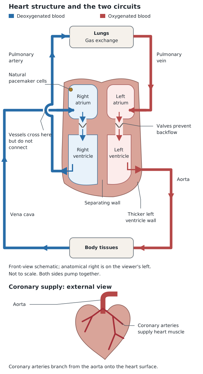
The right ventricle sends blood to the lungs; the left ventricle sends it around the body. Arrows show the route, not the timing: both sides pump together.
The ventricles have thicker muscular walls than the atria because they must generate enough pressure to pump blood out of the heart. The left ventricle has a thicker muscular wall than the right ventricle: its stronger contraction generates the higher pressure needed to send blood around the whole body. The right ventricle pumps blood only to the nearby lungs, at lower pressure.
A wall separates the right and left sides, preventing oxygenated and deoxygenated blood from mixing. Valves prevent backflow, maintaining one-way movement through the heart. Both sides work together; the right and left ventricles contract at the same time.
A group of cells in the right atrium acts as the heart's natural pacemaker. These cells control the natural resting heart rate: the number of times the heart beats each minute while a person is at rest. They produce electrical impulses that cause the heart muscle to contract.
An artificial pacemaker is an electrical device used to correct irregularities in heart rate. It delivers electrical impulses to help the heart beat regularly when its natural rhythm is not working properly.
Air travels along the trachea, or windpipe, which divides into two bronchi, one entering each lung. Within the lungs, the airways lead to many tiny air sacs called alveoli. A network of capillaries surrounds the alveoli, bringing blood close to the air inside them.
Gas exchange occurs by diffusion: the net movement of particles from a higher to a lower concentration. Oxygen moves from the air in the alveoli into the blood, down its concentration gradient. Carbon dioxide moves in the opposite direction, from the blood into the alveoli, and is then breathed out.
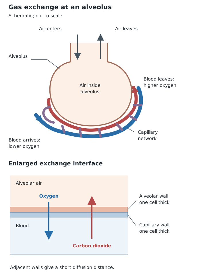
Thin walls give a short diffusion distance. Ventilation and blood flow maintain the gradients for oxygen and carbon dioxide to diffuse in opposite directions.
The lungs contain very many alveoli, giving a large total surface area. This allows more gas to diffuse across the exchange surface at the same time.
The alveolar walls and capillary walls are each one cell thick, so gases travel only a short distance between the air and the blood. A shorter diffusion distance makes exchange faster.
Ventilation continually replaces the air in the alveoli. It keeps the oxygen concentration relatively high and the carbon dioxide concentration relatively low. This maintains the concentration gradients needed for diffusion.
A good blood supply, provided by the dense capillary network, also maintains these gradients. Blood brings carbon dioxide to the alveoli and carries absorbed oxygen away. Ventilation and blood flow therefore work together: one refreshes the air and the other refreshes the blood.
There are three main types of blood vessel. Arteries carry blood away from the heart, veins carry blood towards the heart, and capillaries connect the arterial and venous sides within tissues. Blood travels through the vessel's internal space, called its lumen.
Arteries usually carry blood at high pressure, generated by the contracting heart. Their thick walls contain muscle for strength and elastic tissue that stretches and recoils as blood is pumped through. These walls withstand the pressure and help maintain blood flow between heartbeats. Arteries have a narrower lumen than veins.
Veins return blood at much lower pressure, so their walls are thinner and contain less muscle and elastic tissue. Their wide lumen allows a large volume of blood to flow with little resistance. Valves prevent blood from flowing backwards.
Capillaries are the sites of exchange between blood and tissues. Their walls are only one cell thick, giving a short diffusion distance for substances such as oxygen and carbon dioxide. Their extremely narrow lumen brings blood close to the wall. Extensive capillary networks provide a large area for exchange. In the lungs, these networks surround the alveoli; elsewhere, they supply body cells and remove wastes.
Blood-flow rate describes the volume of blood passing a point in a given time. Calculate it by dividing the volume by the time:
For example, suppose 120 cm³ of blood passes a point in 30 seconds. The mean blood-flow rate is:
This means that an average of 4 cm³ passes the point each second. To express the same rate per minute, multiply by 60: . The volume and time units determine the units of the rate.
You can also rearrange the relationship: volume = rate × time and time = volume ÷ rate. When multiplying a rate by a time, use matching time units: a rate in cm³/min requires a time in minutes.
Get unlimited access to all revision notes, key terms, and exam tips.
Body → vena cava → right atrium → right ventricle → pulmonary artery → lungs → pulmonary vein → left atrium → left ventricle → aorta → body.
Trachea → bronchi → alveoli, surrounded by capillaries.
| Vessel | Direction or role | Structural adaptations |
|---|---|---|
| Artery | Away from heart; usually high pressure | Thick muscular, elastic wall; narrower lumen than a vein. |
| Vein | Towards heart; low pressure | Thinner wall, wide lumen, valves preventing backflow. |
| Capillary | Exchange with tissues | Wall one cell thick; very narrow lumen; extensive networks. |
Volume = rate × time; time = volume ÷ rate. Use matching time units and give rate in units such as cm³/s or cm³/min.
Get unlimited access to all revision notes, key terms, and exam tips.
Define arteries and veins by the direction of blood flow, not by whether the blood is oxygenated.
Link each adaptation to its effect: walls one cell thick shorten the diffusion distance; ventilation and blood flow maintain concentration gradients.
Distinguish ventilation, which moves air, from gas exchange, which occurs by diffusion.
The right ventricle pumps blood to the lungs; the left ventricle pumps blood to the rest of the body.
In a front-view heart diagram, the person's right side appears on the left of the page.
For blood-flow calculations, check the time unit and include a volume-per-time unit in your answer.
You do not need to learn the names of the heart valves.
Double circulatory system
A transport system in which blood passes through the heart twice during one complete circuit: once through the lungs and once through the rest of the body.
Atrium
An upper chamber of the heart that receives blood.
Ventricle
A lower chamber of the heart that pumps blood out of the heart.
Artery
A blood vessel that carries blood away from the heart.
Vein
A blood vessel that carries blood towards the heart.
Capillary
A very small blood vessel where substances are exchanged between blood and surrounding tissues.
Lumen
The internal space of a blood vessel through which blood flows.
Alveolus
A tiny air sac in the lungs where gases are exchanged between air and blood.
Diffusion
The net movement of particles from a region of higher concentration to a region of lower concentration.
Ventilation
The movement of air into and out of the lungs.
Pacemaker
A group of cells in the right atrium that controls the natural resting heart rate.
Artificial pacemaker
An electrical device used to correct irregularities in heart rate.
Put your knowledge into practice — try past paper questions for Combined Science Trilogy
Double circulatory system
A transport system in which blood passes through the heart twice during one complete circuit: once through the lungs and once through the rest of the body.
Atrium
An upper chamber of the heart that receives blood.
Ventricle
A lower chamber of the heart that pumps blood out of the heart.
Artery
A blood vessel that carries blood away from the heart.
Vein
A blood vessel that carries blood towards the heart.
Capillary
A very small blood vessel where substances are exchanged between blood and surrounding tissues.
Lumen
The internal space of a blood vessel through which blood flows.
Alveolus
A tiny air sac in the lungs where gases are exchanged between air and blood.
Diffusion
The net movement of particles from a region of higher concentration to a region of lower concentration.
Ventilation
The movement of air into and out of the lungs.
Pacemaker
A group of cells in the right atrium that controls the natural resting heart rate.
Artificial pacemaker
An electrical device used to correct irregularities in heart rate.