Loading…


Loading…
Loading…
Loading…
AQA GCSE Combined Science Trilogy · 8464
AQA 8464 · 4.1.1.2 Animal and plant cells Check the specification (PDF) (opens in a new tab)
A light microscope uses light and lenses to enlarge a specimen. It allows you to observe the arrangement of cells and identify some of their sub-cellular structures: the smaller structures within each cell. In this required practical, you observe a selection of plant and animal cells, draw and label them, and include a magnification scale.
Cells must be on a glass microscope slide. You can use prepared plant and animal slides, or make a temporary slide. Onion tissue is a useful plant specimen; cheek cells are a common animal specimen. Most cells have little colour, so a stain increases contrast between their structures. Iodine can stain onion cells, while methylene blue can stain cheek cells.
To prepare an onion slide, use tweezers to peel a thin layer of tissue from an onion layer. Place it flat on a slide without folds, and add a drop of iodine. Carefully lower a coverslip at an angle to avoid trapping air bubbles. The coverslip keeps the specimen flat and retains liquid underneath it. Blot away excess liquid with a paper towel. A thin, flat specimen lets light pass through without several layers of cells obscuring each other.
The stage supports the slide, and clips hold it in position. The objective lenses, mounted on a rotating nosepiece, provide different magnifications. You look through the eyepiece lens. Coarse adjustment makes larger focusing movements; fine adjustment makes smaller movements that sharpen the image.
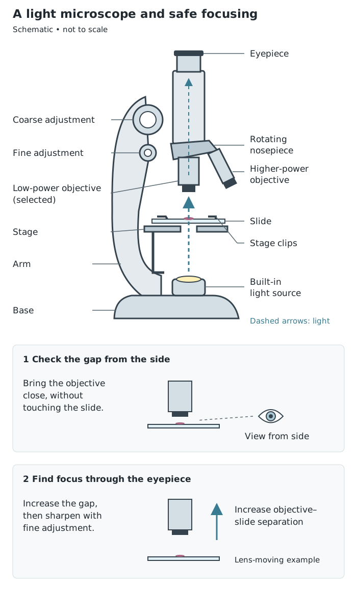
Start with the low-power objective. Watch from the side when bringing it close to the slide, then increase the separation to find the focus.
Use a light microscope, plant and animal slides, and a pencil and ruler for recording your observations.
Higher power enlarges individual cells but shows a smaller area of the specimen. Repeat the observations with both plant and animal specimens.
On an onion slide, look for the cell walls outlining neighbouring cells and, where clearly stained, nuclei and cytoplasm. On a cheek-cell slide, look for each cell's boundary, cytoplasm and stained nucleus. The exact structures visible depend on the specimen, staining and focus. Ribosomes are too small to distinguish with a school light microscope; the absence of visible detail does not mean a structure is absent from the cell.
Make a large, clear pencil drawing of a few representative cells. Use continuous outlines without shading or sketchy strokes, and keep the structures in proportion. Draw straight label lines with a ruler, touching the structures they identify and avoiding crossed lines. Give the drawing a title identifying the specimen.
Record the microscope magnification underneath:
For example, a ×10 eyepiece with a ×4 objective gives ×40 viewing magnification. With a ×40 objective, the same eyepiece gives ×400.
A scale bar gives a direct indication of actual size. Its length must match the scale of the particular image. For example, in an image enlarged ×1000, a 10 mm bar represents 10 µm in the specimen: 10 µm is 0.01 mm, and . Label the bar 10 µm, because that is the actual distance represented. Do not attach an arbitrary scale bar to a drawing whose scale is unknown.
Estimation is useful when you need an approximate comparison rather than an exact measurement. Cell boundaries may be irregular or unclear, and structures vary between cells, so reporting a highly precise value can be misleading.
Compare structures within the same image, or images at the same magnification. Measure the same kind of dimension for both: for example, width across the nucleus and width across the whole cell.
In one image, suppose a cell measures about 24 mm across and its nucleus about 6 mm across. Dividing shows that the cell is about four times as wide as its nucleus. The actual dimensions are not needed for this relative comparison, because both image measurements have been enlarged by the same factor.
Area is the amount of a two-dimensional image occupied by a structure. It is different from length, so a width comparison alone does not establish an area comparison.
One practical way to estimate area is to place a square grid over an image. Count complete squares within each boundary, then combine partly covered squares into approximate whole squares. Use the same grid for both structures.
For one cell image, imagine that the whole cell covers about 40 squares and its nucleus covers about 8. The nucleus occupies about of the cell's image area. If the remaining area is cytoplasm, its estimated area is squares, giving a cytoplasm-to-nucleus area ratio of , or .
These are estimates of the areas visible in the image, not measurements of the structures' three-dimensional volumes. Clearer images and more careful boundary measurements are needed when a precise value matters.
Get unlimited access to all revision notes, key terms, and exam tips.
Record viewing magnification. A scale bar shows an actual specimen distance and must match the image's scale.
Start with the lowest-power objective to locate the cells, then use fine adjustment after switching to higher power.
Draw and label what you can actually see, rather than adding every structure from a textbook cell diagram.
Multiply eyepiece and objective magnifications; do not add them.
Distinguish the microscope's viewing magnification from the magnification of your drawing. A hand-drawn image is not automatically the same size as the microscope image.
Compare lengths with lengths and areas with areas. A structure twice as wide does not necessarily have twice the area.
Use approximate language for estimates and compare structures shown at the same magnification.
Light microscope
A microscope that uses visible light and lenses to produce an enlarged image of a specimen.
Sub-cellular structure
A structure within a cell, such as a nucleus or chloroplast.
Magnification
The number of times larger an image is than the actual object.
Field of view
The area of the specimen visible through a microscope at one time.
Scale bar
A line on an image labelled with the actual distance it represents in the specimen.
Stain
A substance used to add colour and contrast so that cells or their structures are easier to distinguish.
Estimation
An approximate value based on observations or measurements, used when an exact value is unnecessary or cannot be obtained reliably.
Put your knowledge into practice — try past paper questions for Combined Science Trilogy
Light microscope
A microscope that uses visible light and lenses to produce an enlarged image of a specimen.
Sub-cellular structure
A structure within a cell, such as a nucleus or chloroplast.
Magnification
The number of times larger an image is than the actual object.
Field of view
The area of the specimen visible through a microscope at one time.
Scale bar
A line on an image labelled with the actual distance it represents in the specimen.
Stain
A substance used to add colour and contrast so that cells or their structures are easier to distinguish.
Estimation
An approximate value based on observations or measurements, used when an exact value is unnecessary or cannot be obtained reliably.
Get unlimited access to all revision notes, key terms, and exam tips.