

Loading…
Loading…
Loading…
OCR GCSE Psychology · J203
OCR J203 · Development of Treatments Check the specification (PDF) (opens in a new tab)
Neuropsychology investigates how the brain’s structure and functioning relate to behaviour and mental processes. For example, difficulties with concentration or planning can be studied alongside differences in brain activity. This provides a biological approach to understanding schizophrenia and clinical depression.
Researchers use two complementary tools. Neuropsychological tests measure how well someone performs a mental task. Brain imaging reveals physical features of the brain or indicators of its activity. Together, they can connect difficulties in thinking with the brain systems involved, rather than relying only on descriptions of symptoms.
Early understanding depended heavily on people with brain injuries. If damage to a particular area was followed by a loss of an ability, researchers could investigate that area's possible role. Post-mortem examinations also allowed the brain to be compared with records of the person's behaviour during life. These approaches were valuable, but unusual cases were difficult to generalise from, and researchers could not watch the living brain working.
Standardised tests made it possible to assess mental abilities in living people. The Wisconsin Card Sorting Test was published by Grant and Berg in 1948. In the 1960s, Brenda Milner used card sorting to investigate the effects of brain damage, particularly damage involving the frontal lobes. This helped connect measurable performance with brain functioning.
From the 1970s onwards, imaging techniques such as CT added pictures of the living brain. Later functional imaging allowed researchers to investigate activity while people performed tasks. Neuropsychology could therefore study schizophrenia and clinical depression without waiting for brain injury or death. Imaging extended earlier knowledge rather than making tests or injury evidence unnecessary.
A neuropsychological test assesses an ability such as memory, attention or problem-solving. Standardisation means using consistent instructions, procedures and scoring. Researchers can compare someone's performance with typical results, including results for people of a similar age, to identify particular difficulties.
For example, a memory task might involve hearing a list of words and recalling as many as possible. The recall score measures performance on that task; it does not directly show an image of the brain. In clinical depression, testing can reveal poorer concentration, slower thinking and difficulties with memory.
Repeated testing can also help assess treatment. If performance improves over time, this may provide evidence of improvement in cognitive functioning. However, interpreting a score still requires information about the individual and the circumstances of testing.
The Wisconsin Card Sorting Test, or WCST, assesses reasoning and flexible thinking. Participants sort cards containing symbols that differ in colour, shape and number. They are not told the sorting rule: feedback tells them whether their choices are correct, so they must work out the rule.
The rule then changes, requiring a new strategy. For example, suppose matching by colour initially produces correct feedback, but the rule changes to matching by shape. The participant must recognise that the old approach no longer works and adapt. Continuing to sort by colour despite negative feedback is an example of perseveration.
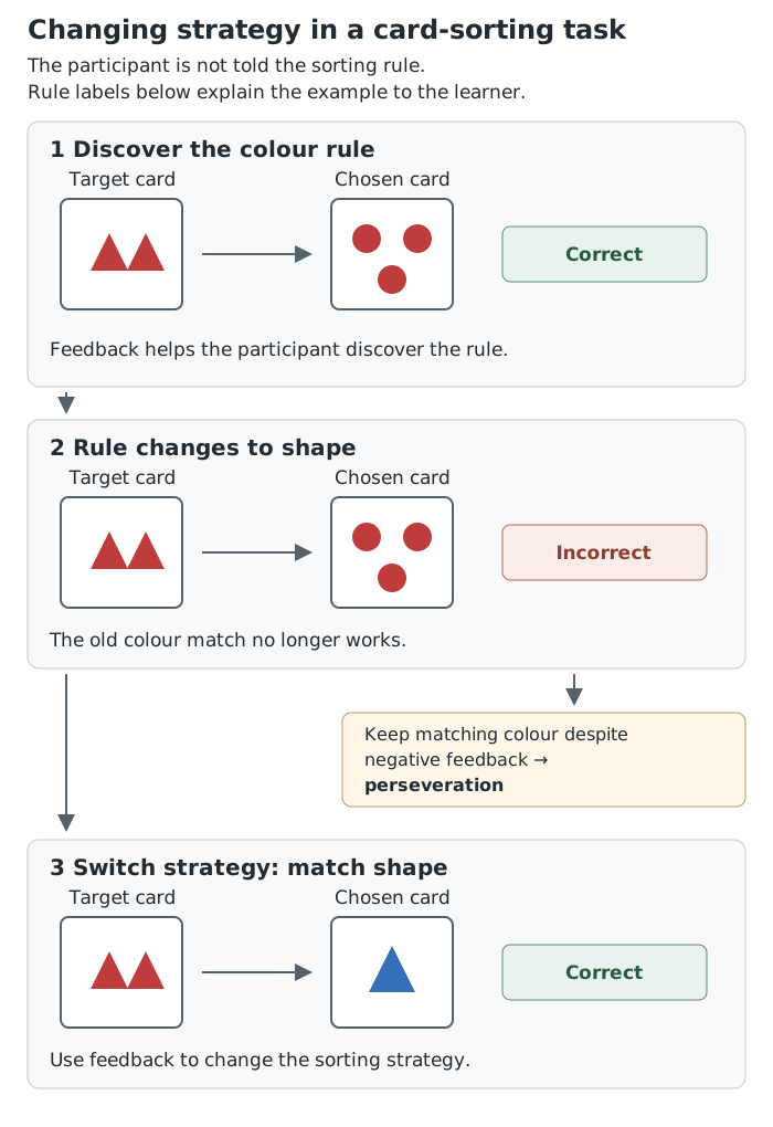
A change in the sorting rule requires the participant to use feedback and switch strategy.
This ability to plan, use feedback and switch strategies is associated with the frontal lobes, at the front of the cerebrum. People with schizophrenia often perform poorly on the WCST. Such performance can suggest difficulties in frontal-lobe functioning, but does not prove that a particular brain area is damaged or establish a diagnosis.
Depression can also be assessed using questionnaires such as the Beck Depression Inventory (BDI). This is a multiple-choice, self-report measure of depressive symptoms, including mood, hopelessness and tiredness. Unlike a card-sorting or memory task, it measures reported symptom severity rather than directly testing a cognitive ability. This distinction matters when interpreting what a score tells us.
Structural scans, including CT and MRI, show the brain's physical features. Researchers can examine its size and shape and look for damage or differences in particular structures. In schizophrenia, some scans show enlarged ventricles, the fluid-filled spaces within the brain, or a smaller hippocampus, a structure involved in memory. Reduced hippocampal volume has also been associated with long-lasting clinical depression.
Functional scans, including fMRI, PET and SPECT, investigate indicators of activity. They do not photograph thoughts. fMRI detects changes associated with blood flow and oxygenation: active brain areas need an increased oxygen supply. PET uses an injected radioactive tracer to investigate processes such as energy use. Researchers use these indicators to compare activity across areas or between groups.
In schizophrenia, research has found reduced frontal-lobe activity, including in the prefrontal region. In clinical depression, imaging can reveal differences in activity in areas involved in mood, including reduced frontal activity. These are patterns found in research, not features present in every person with either condition.
Tests and scans can be used together. Daniel and colleagues (1991) combined the Wisconsin Card Sorting Test with SPECT imaging to investigate prefrontal blood flow. This illustrates how performance on a thinking task can be studied alongside brain functioning.
Tests provide numerical scores, while scans provide measurable information about structure or activity. These methods make comparisons more objective and allow researchers to study living people without brain surgery. They help investigate biological explanations, assess changes during treatment and understand why people may respond differently to treatment. Scans can also help identify other physical problems, such as a tumour.
However, a brain difference associated with a disorder is not necessarily its cause. It might develop as a consequence of the condition or be affected by long-term medication. For example, a smaller hippocampus in someone with years of depression does not show that the smaller hippocampus existed before their depression began.
Findings also overlap: people without a disorder may show similar differences, while people with the disorder may not show them. Tests and scans therefore cannot diagnose schizophrenia or clinical depression on their own. Finally, a purely biological explanation can be reductionist, overlooking psychological experiences and social circumstances. Neuropsychological evidence is most useful when combined with clinical assessment and other explanations.
Value: measurable evidence; study living brains; compare groups; investigate treatment effects.
Limits: association is not causation; medication or the disorder may produce differences; findings overlap between groups; biology alone overlooks psychological and social factors.
Distinguish the methods: a neuropsychological test measures performance on a mental task; brain imaging shows brain structure or indicators of activity.
Name a technique and explain what it measures, rather than simply referring to ‘brain research’.
Use cautious conclusions: an association between a brain difference and a disorder does not establish which caused which.
Neither a poor card-sorting score nor a brain scan diagnoses schizophrenia or clinical depression on its own.
It can see this note and what your specification asks for.
Answers are free with an account.
1 free note left this week
Get more free notesYour answer sets when this note comes back for review, or marks it complete.
Was this note helpful?
Log in to tell us whether it helped, or to report a problem.
Last updated
Neuropsychology
The study of relationships between the brain’s structure and functioning and behaviour or mental processes.
Neuropsychological test
A standardised task used to assess a mental ability, such as memory, attention or problem-solving, to help understand brain functioning.
Standardisation
Keeping the instructions, task and scoring procedures consistent so that people's results can be compared fairly.
Wisconsin Card Sorting Test
A card-sorting task that assesses reasoning and the ability to change strategy when a sorting rule changes; performance is linked to frontal-lobe functioning.
Perseveration
Continuing to use an old response or strategy even when feedback shows that it is no longer appropriate.
Structural brain imaging
A technique that produces images of the brain’s physical features, such as its size, shape and internal structures.
Functional brain imaging
A technique that maps indicators of brain activity, such as changes in blood flow or energy use.
Put your knowledge into practice — try past paper questions for Psychology
Neuropsychology
The study of relationships between the brain’s structure and functioning and behaviour or mental processes.
Neuropsychological test
A standardised task used to assess a mental ability, such as memory, attention or problem-solving, to help understand brain functioning.
Standardisation
Keeping the instructions, task and scoring procedures consistent so that people's results can be compared fairly.
Wisconsin Card Sorting Test
A card-sorting task that assesses reasoning and the ability to change strategy when a sorting rule changes; performance is linked to frontal-lobe functioning.
Perseveration
Continuing to use an old response or strategy even when feedback shows that it is no longer appropriate.
Structural brain imaging
A technique that produces images of the brain’s physical features, such as its size, shape and internal structures.
Functional brain imaging
A technique that maps indicators of brain activity, such as changes in blood flow or energy use.