

Loading…
Loading…
Loading…
AQA GCSE Combined Science: Synergy · 8465
AQA 8465 · 4.1.3.1 Check the specification (PDF) (opens in a new tab)
Cells contain smaller parts called sub-cellular structures, such as nuclei and mitochondria. Microscopes allow us to study structures that are too small to examine with the unaided eye.
A light microscope uses light and lenses to form a magnified image. Light microscopes can show cells and larger sub-cellular structures, including nuclei and vacuoles. Their development from the seventeenth century allowed scientists to explore the cellular organisation of living things.
Magnification describes how many times larger an image is than the real object. Resolving power describes the ability to distinguish two points close together as separate points. These are different properties: enlarging a blurred image makes the blur bigger, but does not reveal the missing detail.
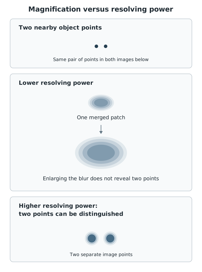
A larger image does not necessarily show more detail. Higher resolving power allows closely spaced points to be seen separately.
Electron microscopes were introduced in the twentieth century. They use a beam of electrons rather than light and have much higher resolving power than light microscopes. Closely spaced features that merge together in a light-microscope image can therefore be distinguished using an electron microscope.
A light microscope has a useful magnification of about ×1000. An electron microscope can magnify ×1 000 000 or more. Its higher resolving power makes this much greater magnification useful: scientists can study cells in finer detail rather than simply viewing a larger blur.
This has increased our understanding of sub-cellular structures. For example, electron microscopy allows finer examination of mitochondria and has improved our understanding of the structure of the nucleus and cell membrane. The important connection is higher resolving power → finer structural detail → better understanding of cells.
Microscopic objects are often described in micrometres or nanometres. The prefixes tell you the fraction of a metre represented by each unit.
| Prefix | Unit | Size in metres |
|---|---|---|
| centi | centimetre, cm | m |
| milli | millimetre, mm | m |
| micro | micrometre, µm | m |
The useful conversion relationships are:
When converting to a smaller unit, the numerical value increases because more of those units fit into the same length. For example, . To convert in the opposite direction, divide by the appropriate factor.
Magnification compares an image length with the corresponding length of the real object:
Writing magnification as , image size as and real size as gives three useful forms:
Both lengths must use the same unit. Their units cancel when calculating magnification, so magnification is written as, for example, ×500, not 500 mm.
Suppose a cell is long and its image is long. Convert the image length to , then calculate . The image is 500 times as long as the real cell.
To find a real size, suppose a structure's image measures at ×15 000. Its real size is , or .
To find an image size, suppose an object is long and is magnified ×2000. Its image length is , or . Multiplication makes sense here because the image is larger than the object.
Standard form writes a number as , with . It keeps very small lengths and very large manageable. For example, .
Suppose an image length is and the corresponding real length is . The units already match:
Dividing the coefficients gives 3.0; subtracting the powers gives . The magnification is ×3000, expressed as to retain two significant figures.
Count significant figures from the first non-zero digit. For example, has three significant figures and rounds to to two significant figures because the next digit is 8. Use the precision requested in a question; otherwise, choose a precision appropriate to the measurements rather than copying every calculator digit.
An estimate is an approximate answer. It is useful before a calculation to predict the scale of the result, afterwards to check its plausibility, or when measurements themselves are only approximate. One useful method is to round quantities to one significant figure before calculating.
For example, suppose an image length is and the real object length is . Approximating these as and gives an estimated magnification of ×2000. An exact calculation should be close to this; a very different answer suggests checking the units or arithmetic.
Orders of magnitude compare sizes using powers of ten. Suppose two objects have lengths of and . Their size ratio is : the larger is 1000 times as long, a difference of . Convert to the same unit before making this comparison.
For the benefit of electron microscopy, link higher resolving power to finer visible detail and better understanding of sub-cellular structures—not just a larger image.
Convert image size and real size to the same unit before calculating magnification. Magnification has no unit.
Check whether the question asks for magnification, real size or image size, and rearrange the equation accordingly.
Keep calculator precision during a calculation, then round the final answer to the requested number of significant figures. Round directly, not in successive stages.
It can see this note and what your specification asks for.
Answers are free with an account.
1 free note left this week
Get more free notesYour answer sets when this note comes back for review, or marks it complete.
Was this note helpful?
Log in to tell us whether it helped, or to report a problem.
Last updated
Sub-cellular structure
A structure within a cell, such as a nucleus, mitochondrion or ribosome.
Magnification
How many times larger an image is than the real object: .
Resolving power
The ability to distinguish two points close together as separate points. Higher resolving power allows finer detail to be seen.
Electron microscope
A microscope that uses a beam of electrons to produce an image and has much higher resolving power than a light microscope.
Light microscope
A microscope that uses light and lenses to produce a magnified image.
Standard form
A way of writing a number as , where and is an integer.
Order of magnitude
A scale of size expressed using powers of ten. A difference of one order of magnitude means a factor of ten.
Significant figure
A digit counted when expressing the precision of a number, starting with its first non-zero digit.
Put your knowledge into practice — try past paper questions for Combined Science: Synergy
Sub-cellular structure
A structure within a cell, such as a nucleus, mitochondrion or ribosome.
Magnification
How many times larger an image is than the real object: .
Resolving power
The ability to distinguish two points close together as separate points. Higher resolving power allows finer detail to be seen.
Electron microscope
A microscope that uses a beam of electrons to produce an image and has much higher resolving power than a light microscope.
Light microscope
A microscope that uses light and lenses to produce a magnified image.
Standard form
A way of writing a number as , where and is an integer.
Order of magnitude
A scale of size expressed using powers of ten. A difference of one order of magnitude means a factor of ten.
Significant figure
A digit counted when expressing the precision of a number, starting with its first non-zero digit.
| nano | nanometre, nm | m |
Use matching length units. Magnification has no unit.